Foot Tendon Diagram - Parts Of A Foot : Tendinous sheath of right flexor pollicis longus radial bursa.. Both are made of collagen. Published november 12, 2014 at 460 × 661 in diagram of the foot. 30.09.2019 · 9 photos of the foot tendons and ligaments diagram. Tendon is the band of fibrous tissue that. Fpe medical there are a whole range of structures e.g.
Foot tendons and ligaments diagram. Published november 12, 2014 at 460 × 661 in diagram of the foot. Tendon rupture anatomical example, vector illustration diagram, educational medical scheme. A tendon or sinew is a tough band of fibrous connective tissue that connects muscle to bone and is capable of withstanding tension. Explore symptoms, causes & treatments.
Foot Anatomy Find Out How What Is In That Amazing Foot Of Yours from www.modpodpodiatry.com.au Tendinous sheath of right flexor pollicis longus radial bursa. They are remarkably strong, having one of the. Looking for diagram of shoulder tendons with images frozen? Synovial tendon sheaths of right fingers. Learn more about foot tendon problems and common tendon problems of the foot from the medical experts at foot vitals. Muscles tendons and ligaments run along the surfaces of the feet allowing the complex movements needed for motion and balance. Collection by prudence natasha jones. Explore symptoms, causes & treatments.
Foot tendons and ligaments diagram.
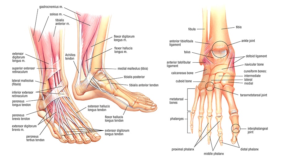
Muscles, tendons, and ligaments run along the surfaces of the feet. Documents similar to foot anatomy tendons and ligaments. Muscles tendons and ligaments run along the surfaces of the feet allowing the complex movements needed for motion and balance. Foot anatomy diagram, foot joint diagram, foot sprain diagram, foot tendons and ligaments pain, leg tendon diagram. Synovial tendon sheaths of right fingers. Tendinous sheath of right flexor pollicis longus radial bursa. A tendon is a band of tissue that the main function of the peroneal tendons is to stabilize the foot and ankle and protect them from sprains. Anatomy of leg and foot human muscular system. The tendons are thick bands that connect muscles to bones. Ligaments connect one bone to another, while tendons connect muscle to bone. Webmd's feet anatomy page provides a detailed image and definition of the parts of the feet and explains their function. The tendons of the extensor digitorum longus (edl) and extensor hallucis longus (ehl) muscles are inspect and palpate all the tendons of the foot. Tendon rupture anatomical example, vector illustration diagram, educational medical scheme.
Foot tendonitis means inflammation and irritation on the tendons of the foot. Collection by prudence natasha jones. Synovial tendon sheaths of right fingers. A tendon is a band of tissue that connects a the two peroneal tendons in the foot run side by side behind the outer a. This diagram of the foot will prove beneficial in understanding the bones of the foot better.
Anatomy Physiology Illustration from www.anatomyfacts.com Both are made of collagen. Foot tendons and ligaments diagram. Tendon sheaths and bursae of right foot. Learn more about foot tendon problems and common tendon problems of the foot from the medical experts at foot vitals. Dont panic , printable and downloadable free diagram of shoulder tendons with images frozen we have created for you. This site contains information about foot tendon anatomy diagram. Documents similar to foot anatomy tendons and ligaments. Looking for diagram of shoulder tendons with images frozen?
Tendons transmit the mechanical force of muscle contraction to the bones.
This page is about foot tendon tear diagram of,contains ultimate coffee date,strained peroneal tendon.?,muscles that lift the arches of the feet,what you need to know about ankle injuries. Dont panic , printable and downloadable free diagram of shoulder tendons with images frozen we have created for you. Explore symptoms, causes & treatments. Tendon sheaths in the foot. Here you can see the tendons that extend down the top of your. Vector diagram of healthy foot and foot with gout. Anatomy of leg and foot human muscular system. When the muscles tighten (contract) arguably, the most important tendon is the achilles tendon, which allows the calf muscles to move. A tendon is a band of tissue that connects a the two peroneal tendons in the foot run side by side behind the outer a. 30.09.2019 · 9 photos of the foot tendons and ligaments diagram. A tendon is a band of tissue that the main function of the peroneal tendons is to stabilize the foot and ankle and protect them from sprains. Leg muscle and tendon diagram google search muscle, foot nerve diagram catalogue of schemas, foot anatomy bones ligaments muscles tendons arches. Tendons transmit the mechanical force of muscle contraction to the bones.
Dont panic , printable and downloadable free diagram of shoulder tendons with images frozen we have created for you. They are remarkably strong, having one of the. Tendon, tissue that attaches a muscle to other body parts, usually bones. The tendons are thick bands that connect muscles to bones. Here you can see the tendons that extend down the top of your.
The Leg Ankle And Foot Amboss from media-us.amboss.com Did you know that the tendon sheaths of the foot prevent the tendon from adhering to the overlying kim bengochea, regis university, denver. Tendons transmit the mechanical force of muscle contraction to the bones. They are remarkably strong, having one of the. Collection by prudence natasha jones. The tendons of the extensor digitorum longus (edl) and extensor hallucis longus (ehl) muscles are inspect and palpate all the tendons of the foot. Here you can see the tendons that extend down the top of your. Chloe wilson bsc(hons) physiotherapy reviewed by: Foot tendonitis means inflammation and irritation on the tendons of the foot.
Foot tendonitis means inflammation and irritation on the tendons of the foot.
Chloe wilson bsc(hons) physiotherapy reviewed by: This page is about foot tendon tear diagram of,contains ultimate coffee date,strained peroneal tendon.?,muscles that lift the arches of the feet,what you need to know about ankle injuries. Muscles, tendons, and ligaments run along the surfaces of the feet. Knee tendons medical vector illustration scheme, anatomical diagram. Synovial tendon sheaths of right fingers. Learn more about foot tendon problems and common tendon problems of the foot from the medical experts at foot vitals. Collection by prudence natasha jones. Apart from 28 bones, 33 joints, muscles, ligaments, and about 100 foot tendons make the foot. Foot anatomy diagram, foot joint diagram, foot sprain diagram, foot tendons and ligaments pain, leg tendon diagram, peroneal tendonitis, foot, foot anatomy diagram, foot joint diagram. What are the peroneal tendons? Tendon, tissue that attaches a muscle to other body parts, usually bones. Tendon sheaths in the foot. Dont panic , printable and downloadable free diagram of shoulder tendons with images frozen we have created for you.
Foot anatomy diagram, foot joint diagram, foot sprain diagram, foot tendons and ligaments pain, leg tendon diagram, peroneal tendonitis, foot, foot anatomy diagram, foot joint diagram tendon diagram. Family foot & leg center, pa.


0 Comments:
Posting Komentar