Home
Uncategories
Groin Female Hip Muscle Anatomy / Adductor Tendinopathy / The groin canal (inguinal canal) connects the inside with the outside of the abdomen and is an opening in the stomach muscles that contains the spermatic cord.
Groin Female Hip Muscle Anatomy / Adductor Tendinopathy / The groin canal (inguinal canal) connects the inside with the outside of the abdomen and is an opening in the stomach muscles that contains the spermatic cord.
Groin Female Hip Muscle Anatomy / Adductor Tendinopathy / The groin canal (inguinal canal) connects the inside with the outside of the abdomen and is an opening in the stomach muscles that contains the spermatic cord.. The canal is like a tube with 4 sides: Adductor magnus is the largest groin muscle and is one of the two long adductor muscles (gracilis is the other). The adductor muscle group, also known as the groin muscles, is a group located on the medial side of the thigh. The psoas major originates from the outer surface of the lower lumbar vertebrae and the iliacus muscle originates from the iliac fossa of the pelvis. Groin and adductors anatomy the groin canal inguinal canal connects the inside with the outside of the abdomen and is an opening in the stomach muscles that contains the spermatic cord.
The groin area sits between the lower part of the stomach, the upper inner thigh, and the hip joint. Joint, soft tissue, bone, back referred, peripheral nerve and other. The psoas major originates from the outer surface of the lower lumbar vertebrae and the iliacus muscle originates from the iliac fossa of the pelvis. These include the liver, stomach, and intestines. The straight line connecting the anterior and posterior margin of the labrum at the midsection of the femur head is located.
Causes, Symptoms and Treatments for Inner Thigh Pain from naturalremedyideas.com Groin and adductors anatomy the groin canal inguinal canal connects the inside with the outside of the abdomen and is an opening in the stomach muscles that contains the spermatic cord. What are calf muscle, skeletal muscle, leg a d foot muscle, hip and groin muscle, shoulder & arm muscles. It also supports the nerves and blood vessels of the leg as they pass through the groin, including the femoral artery, femoral vein, and femoral nerve. Anatomy of the abdomen and groin. These muscles include the gluteus. There is an obvious bump (cam) on the anterior femoral neck (big arrow) with alpha angle over 50° (striped curved line).the acetabulum is overcovering the femoral head (small arrows); The iliopsoas is a muscle formed from the psoas major muscle and the iliacus muscle. The groin is the area in the body where the upper thighs meet the lowest part of the abdomen.
Each of these tissues is discussed in the tabs listed below:
The groin canal (inguinal canal) connects the inside with the outside of the abdomen and is an opening in the stomach muscles that contains the spermatic cord. Adductor magnus is the largest groin muscle and is one of the two long adductor muscles (gracilis is the other). Groin and adductors anatomy the groin canal inguinal canal connects the inside with the outside of the abdomen and is an opening in the stomach muscles that contains the spermatic cord. Groin strain, also referred to as a pulled groin muscle, typically occurs as a result of an athletic injury or awkward movement of the hip joint, which leads to stretching or tearing of the inner thigh muscles. Hip adductor muscles together make up the groin area. Musculoskeletal issues begin in the bones,. Muscle anatomy body anatomy hip muscles anatomy hip anatomy human anatomy pelvis anatomy thigh muscles soft tissue injury psoas release. It also supports the nerves and blood vessels of the leg as they pass through the groin, including the femoral artery, femoral vein, and femoral nerve. These groin muscles adduct the thigh (bring the femur and knee closer to the midline). Chapter outline introduction intraarticular hip pathology joint effusion synovitis proliferative synovial disorders acetabular labrum extraarticular hip pathology muscle and tendon disorders hernias soft tissue masses inguinal lymphadenopathy groin abscess vascular lesions masses in the female groin compression neuropathy hip prosthesis introduction the hip region is an area of complex anatomy. The groin is the area that lies between the abdomen stomach and thighs. These muscles move the thigh toward the body's midline. The different anatomical areas of the gluteal region:
Because the anterior, or front, of your hip and your groin. Musculoskeletal issues begin in the bones,. These include the liver, stomach, and intestines. Hip pain explained will teach you about the anatomy of the hips and pelvic area and how many different types of body tissues interact. The groin is the area in the body where the upper thighs meet the lowest part of the abdomen.
Did you know: Muscles might affect how you feel and your health? - Rodger Duckworth Physiotherapy from rdphysio.com The groin area sits between the lower part of the stomach, the upper inner thigh, and the hip joint. Your groin is the area where your upper thigh and lower abdomen meet. Your hip joint is found along the same line underneath your groin. Anatomy of the groin area superficial muscles and deep muscles in this image, you will find rectus abdominis, external oblique, inguinal ligament, tensor fascia lata, gracilis, sartorius, rectus femoris, the iliotibial band in it. There is an obvious bump (cam) on the anterior femoral neck (big arrow) with alpha angle over 50° (striped curved line).the acetabulum is overcovering the femoral head (small arrows); These include the liver, stomach, and intestines. This issue occurs when extra bone growth on either the acetabulum or femoral head portion of the hip causes the joint to take on an irregular shape. Groin strain, also referred to as a pulled groin muscle, typically occurs as a result of an athletic injury or awkward movement of the hip joint, which leads to stretching or tearing of the inner thigh muscles.
The different anatomical areas of the gluteal region:
Straining adductor muscles of the hip will result in pulled groin muscles which is painful. The different anatomical areas of the gluteal region: Front layer (external oblique fascia), back layer (posterior wall), medial layer (straight abdominal muscles) and lateral layer (inguinal ligament). It lies between the chest and the pelvis, holding many of the body's organs. Hip pain explained will teach you about the anatomy of the hips and pelvic area and how many different types of body tissues interact. Anatomy of the groin area superficial muscles and deep muscles in this image, you will find rectus abdominis, external oblique, inguinal ligament, tensor fascia lata, gracilis, sartorius, rectus femoris, the iliotibial band in it. Causes of pain in the hip and groin can be musculoskeletal or internal. According to wikipedia, the groin area is the junction area between the abdomen and the thighs. It is usually described as having two parts, hamstring and adductor parts. The region has pubic bones which consist of adductor muscles of the hip or the groin muscles. These muscles move the thigh toward the body's midline. Pulled groin muscle these hip adductor muscles that make up the groin consist of the adductor brevis, adductor longus, adductor magnus, gracilis, and pectineus. These muscles include the gluteus.
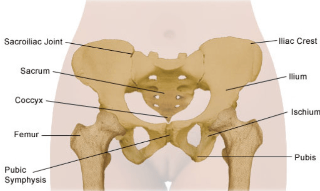
The abdomen is the largest space (cavity) in the body. The adductor muscle group, also known as the groin muscles, is a group located on the medial side of the thigh. These include the liver, stomach, and intestines. The ilium, pubis, and ischium of each hip bone come together to form the acetabulum, where the head of the thigh bone (femur) attaches. Causes of pain in the hip and groin can be musculoskeletal or internal.
Muscles Of Floor Anatomy - Carpet Vidalondon from i.ytimg.com Pulled groin muscle these hip adductor muscles that make up the groin consist of the adductor brevis, adductor longus, adductor magnus, gracilis, and pectineus. These groin muscles adduct the thigh (bring the femur and knee closer to the midline). Joint, soft tissue, bone, back referred, peripheral nerve and other. Your hip joint is found along the same line underneath your groin. Inferior ramus of pubis and ischial ramus. Your groin is the area where your upper thigh and lower abdomen meet. The straight line connecting the anterior and posterior margin of the labrum at the midsection of the femur head is located. The groin region consists of ligaments, tendons, muscles and fascia all of which attach to the pubic bone.
In this video i have explained the iliopsoas muscle and the anatomy of the hip.
I focused on explaining the origin of psoas major and psoas minor as well as. The inguinal ligament supports the muscles that run inferior to its fibers, including the iliopsoas and pectineus muscles of the hip. There is an obvious bump (cam) on the anterior femoral neck (big arrow) with alpha angle over 50° (striped curved line).the acetabulum is overcovering the femoral head (small arrows); The femur, the hip bone (subdivided into ilium, ischium and pubis) as well as the sacrum were labeled separately with differently colored labels In this video i have explained the iliopsoas muscle and the anatomy of the hip. In human anatomy, the groin is the junctional area between the the groin muscles consist of three large groups of muscles that can be injured: The ilium, pubis, and ischium of each hip bone come together to form the acetabulum, where the head of the thigh bone (femur) attaches. Included in this group are the adductor longus, adductor brevis, adductor magnus, pectineus, and gracilis muscles. The groin is the area in the body where the upper thighs meet the lowest part of the abdomen. It also supports the nerves and blood vessels of the leg as they pass through the groin, including the femoral artery, femoral vein, and femoral nerve. Your hip joint is found along the same line underneath your groin. Joint, soft tissue, bone, back referred, peripheral nerve and other. This issue occurs when extra bone growth on either the acetabulum or femoral head portion of the hip causes the joint to take on an irregular shape.
These two muscles joint each other and travel down to form the iliopsoas tendon groin muscle anatomy. These two muscles joint each other and travel down to form the iliopsoas tendon.


0 Comments:
Posting Komentar